An explanation of thoracoscopy (Krames Patient Education)
[Worldtrippers
home] [Mountaintop
home]
[July 19, 2011: All about pneumothorax]
An explanation of thoracoscopy (Krames Patient Education)
After Joss’ thoracoscopy surgery, he had two drain tubes in his left side. The fatter tube drained from his pleural cavity, while the thinner tube drained from his abdomen.
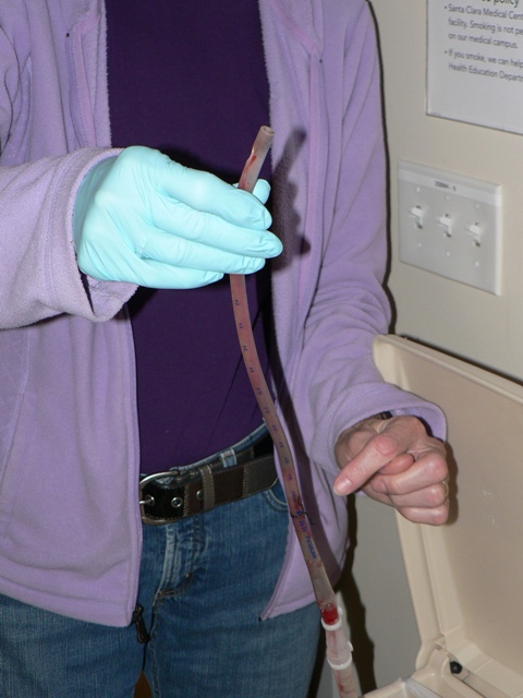
Joss’ pleural catheter was removed after five days. After removing the sutures, a surgical resident simply yanked the entire tube out with one quick pull. Almost 12 inches of tubing had been in Joss’ pleural cavity. (Gail is pointing to the spot where the tube exited Joss’ body.)
Joss’ abdominal catheter was removed after six days. While the exposed part of the tube was thin, the section in his abdomen (white) was actually much fatter. The bulb at the end had provided additional suction for drainage.
Dr. Bloom provided us with this photograph taken with the catheter camera during Joss’ surgery. The ribcage is at the top; the lung is at the bottom. (The instrument at the bottom edge was used to pull the lung away from the ribs.) The white spot in the lower right is the “bleb” on Joss’ left lung. According to Dr. Bloom, the bleb was about 2 centimeters in size.
[Worldtrippers
home] [Mountaintop
home]
[July 19, 2011: All about pneumothorax]